The Mammalian Heart
- The Heart is the organ that controls the circulatory system in mammals (and other animals). It pumps blood around the body. Mammals have a double circulatory system, so the heart must pump blood to the lung and to the rest of body simultaneously.
The Structure of the Heart
On the outside, the heart mainly consists of a dark red muscle. It is attached to four very important blood vessels: the Vena Cava, the Pulmonary Artery, the Pulmonary Vein and the Aorta.
Internally, the heart is made up of four main cavities: two Atria (singular: atrium) and two Ventricles. The atria hold blood briefly, then allow it to fall into the ventricles, which provide the actual ‘pump’.
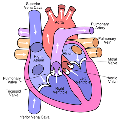
The vena cava supplies de-oxygenated blood from the body, which then flows into the right atrium then the right ventricle. This gets pumped through the pulmonary artery to the lungs where it gets oxygenated, before returning to the heart via the pulmonary vein. This flows through the left atrium into the left ventricle, and then gets pumped to the body via the aorta. It finally returns to the heart through the vena cava, and the process repeats. This is happening inside you right now, about once a second!
The atria are separated from the ventricles by Atrioventricular Valves (specifically called Tricuspid Valves - right; and Bicuspid/Mitral Valves - left). These valves allow blood to flow downwards when the atria and ventricles relax, but close to prevent blood from flowing back up to the atria when the ventricles are contracting.
The ventricles are separated from the aorta and the pulmonary artery by the Semilunar Valves (specifically called, respectively, the Aortic and Pulmonary Valves). These prevent blood from flowing in the wrong direction back into the heart.
The atrial walls are thin; they don’t need to withstand much pressure. The ventricles walls on the other hand are much thicker. When the ventricles contract, the blood pressure inside becomes very high, and they need to be able to withstand this.
Also, the walls of the left ventricle are thicker than those of the right ventricle. This is because the left side of the heart controls the systemic circuit (blood to the whole body) while the right side controls the pulmonary circuit (blood to the lungs).
Blood in the systemic circuit needs to be at a high pressure in order to make its way around the whole body and back again. In contrast, the lungs are very close to the heart, and contain very delicate capillaries which would break if subjected to too great a pressure. Hence the systemic circuit requires a greater blood pressure than the pulmonary circuit, and thus the walls of the left ventricle must be thicker than those of the right ventricle.
The Cardiac Cycle
- The cycle of the beating of the heart consists of three main phases: Diastole, Atrial Systole and Ventricular Systole. The heart cycles through each of those phases in order.

Diastole
During this phase, the heart is filled with blood. The atria and ventricles are relaxing, and blood flows from the major veins (the vena cava and the pulmonary veins) into the atria, then into the ventricles via the open atrioventricular valves.
The atrioventricular valves are open because the pressure in the atria is greater than that in the ventricles. The semilunar valves, on the other hand, are closed because the pressure in the ventricles is lower than that in the main arteries leading from the heart.
Atrial Systole
- This is the beginning of the muscle contraction. The atria contract, which pushes more blood into the ventricles.
Ventricular Systole
During this phase, the ventricles contract, increasing the ventricular pressure. Blood pushes against the atrioventricular valves, since the pressure in the ventricles is now greater than that in the atria, causing them to snap shut, which prevents blood from flowing back into the atria. This snapping shut the the audible ‘lub’ in the familiar ‘lub-dub’ sound of the heart.
The pressure in the ventricles continues to increase until it is greater than that in the main arteries leading from the heart. At this point, the semilunar valves are forced open, and blood rushes out of the ventricles out of the heart into the arteries.
After the ventricles have finished contracting, the muscles relax, and are pulled back by elastic tissue. This decreases the pressure in the ventricles, causing the semilunar valves to shut (the ‘dub’ in the ‘lub-dub’ of the heart) and the atrioventricular valves to open, so that the diastole phase can proceed once more.
Coordination in the Cardiac Cycle
There is a need to control and coordinate the cardiac cycle described above. The heart rate needs to change to respond to various factors, such as increased physical activity. Also, the atria and the ventricles have different natural frequencies of contraction. If the contractions where uncoordinated, the atria and the ventricles would contract asynchronously, which would lead to inefficient pumping.
Heart muscle is Myogenic: it initiates its own contraction. This unusual property means that the heart is able to beat even when not connected to the body.
The Process of a Heartbeat
- A heartbeat begins in a region of tissue known as the Sinoatrial Node. This is located above the right atrium. It generates electrical pulses in a regular fashion, which then spread throughout the heart.

Once an electrical excitation has been generated by the sinoatrial node, it spreads throughout the atrial muscles, which then contact as part of atrial systole.
The wave of excitation is stopped however by a non-conducting disc of tissue at the base of the atria. This prevents the wave from causing the ventricles to contract too early. The only path to the ventricles is through another node called the Atrioventricular Node.
At the atrioventricular node, the electrical pulse is delayed to allow the atria to fully contract. Then the wave travels down the inter-ventricular wall through special conducting tissue known as the Purkyne, towards the apex (base) and the ventricular muscles.
As the wave reaches the muscles, it initiates the contraction. The wave then moves up the ventricles from the apex causing further contraction. In this manner the contraction of the ventricles starts at the bottom and moves upwards, so that blood is forced upwards into the arteries during ventricular systole.
Electrocardiograms
- An Electrocardiogram (ECG) is a device that measures the electrical activity of the heart. It consists of several sensors placed around the body connected to a monitor. The sensors detect the electrical signals that spread from the heart through the body to the skin.

- The ECG produces a trace of the electrical activity. The trace of a healthy person has what’s know as a ‘PQRST shape’. Its waveforms consist of regions labelled by the letters P, Q, R, S and T.

- The wave labelled ‘P’ corresponds to the excitation and contraction of the atria during atrial systole. The ‘QRS’ comlpex corresponds to the excitation and contraction of the ventricles during ventricular systole. Finally, the ‘T’ wave corresponds the diastole phase.